A paper recently published in the journal Light: Science and Applications investigated the effectiveness of microtoroid optical resonator photothermal microscopy for individual 5 nm quantum dot detection.
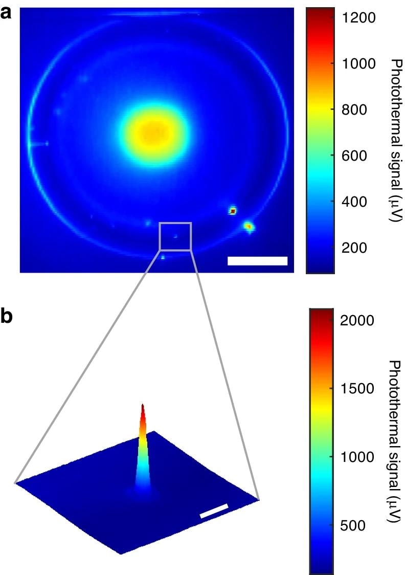
 A coarse photothermal map of the whole microtoroid. The diameter of Au nanosphere on the microtoroid is 100 nm. Scale bar, 20 µm. b Fine photothermal map of single 100 nm Au nanospheres marked in (a). Scale bar, 2 µm. The 2D photothermal maps are generated by scanning the selected area line-by-line using the pump laser. Image Credit: https://www.nature.com/articles/s41377-024-01536-9
Importance of Photothermal Microscopy
Label-free individual molecule and particle detection techniques are critical in nanomaterial investigations, disease diagnostics, and basic science. Although conventional fluorescence-based methods for single-molecule imaging and detection are used for studying molecular processes due to their high sensitivity and low background noise, their effectiveness is limited by photobleaching, photoblinking, and available molecular probes.
Photothermal microscopy has gained significant attention as a noninvasive, label-free imaging technique that can detect single nano absorbers with high sensitivity. Photothermal heterodyne imaging (PHI) is the commonly used technique for photothermal microscopy of individual nanoobjects.
Whispering gallery mode (WGM) microresonators are a suitable ultra-sensitive platform for photothermal microscopy, as they can enhance light-matter interaction by confining light in a small volume. Studies have combined microtoroid optical resonators and photothermal microscopy to detect 100 nm-long polymers and 250 nm-long gold nanorods.
The Study
In this study, researchers combined microtoroids with photothermal microscopy to spatially detect single 5 nm diameter quantum dots with a high signal-to-noise ratio. WGM microtoroid resonators were used as detectors in photothermal microscopy to perform ultra-sensitive photothermal imaging at room temperature with a simpler alignment requirement than PHI.
The objective was to demonstrate that frequency-locked optical whispering evanescent resonator (FLOWER)-based photothermal microscopy using re-etched microtoroids could detect individual nanoparticles, as small as 5 nm quantum dots, with high sensitivity.
FLOWER integrates optical microcavities with data processing and frequency locking to enable single macromolecule detection. Traditional approaches involve scanning a tunable probe laser’s wavelength, where the measuring time and resonance measurement accuracy are constrained by the laser scanning speed and compromised by laser drift, respectively.
However, FLOWER utilizes frequency locking to improve the resonance shift measurement accuracy and decrease the response time. The pump laser’s point-by-point scanning generated photothermal images.
Experimental Setup: In the experimental setup, a probe laser with 780 nm wavelength was coupled into the microtoroid resonator using a tapered optical fiber. A polarization controller was employed to control the probe laser’s polarization to optimize the coupling efficiency between the microtoroid and tapered fiber.
Additionally, a 25 MHz oscillation dither signal was applied to drive the phase modulator, leading to the probe laser’s phase modulation. A fiber-coupled beam splitter was utilized to split the phase-modulated probe laser into two arms, with one arm serving as the reference light, while the other carrying the signal light coupled into the microtoroid.
Subsequently, both the reference and signal light were received by a balanced high-bandwidth photodetector, which played a critical role in eliminating common noise from the probe laser. By multiplying the electrical output signal of the balanced photodiode with the time-averaging and dither signal, an error signal proportional to the wavelength detuning was obtained between the microtoroid’s WGM resonance and the probe laser.
The proportional-integral-derivative controller used this error signal in a feedback loop with the tunable laser controller to minimize the error signal, ensuring stable and accurate probe laser’s frequency locking to the WGM resonance.
Importance of this Work
Researchers successfully displayed the single nanoparticle/quantum dot detection ability of the FLOWER-based photothermal microscopy using re-etched microtoroids. 5 nm diameter quantum dots were spatially detected with over 104 signal-to-noise ratio.
High-sensitivity fluorescence imaging confirmed single particle detection for 18 nm quantum dots and for 5 nm quantum dots through comparison with theory. The proposed system demonstrated the ability to detect a minimum heat dissipation of 0.75 pW.
Specifically, the 0.75 pW detection limit of photothermal microscopy in heat dissipation was over two orders of magnitude smaller compared to the 0.34 nW heat dissipation power previously observed for single dye molecules during photothermal detection. This highlighted the proposed photothermal microscopy's ability to detect single molecules. The significant advancement of the detection limit was attributed to the improvement in photothermal sensitivity.
In conclusion, this study's findings demonstrated the feasibility of using microtoroid optical resonator photothermal microscopy for single 5-nm quantum dot detection.
Thus, this system could be effectively used in several fields, including medicine, chemistry, materials science, nanotechnology, and biological sciences. In the future, this system can be integrated with specific capture agents like sorbent polymers or aptamers to offer enhanced specificity for many applications.
Journal Reference
Hao, S., Suebka, S., Su, J. (2024). Single 5-nm quantum dot detection via microtoroid optical resonator photothermal microscopy. Light: Science & Applications, 13(1), 1-11. DOI: 10.1038/s41377-024-01536-9, https://www.nature.com/articles/s41377-024-01536-9
Disclaimer: The views expressed here are those of the author expressed in their private capacity and do not necessarily represent the views of AZoM.com Limited T/A AZoNetwork the owner and operator of this website. This disclaimer forms part of the Terms and conditions of use of this website.