Jul 25 2016
A novel electron microscope capable of visualizing electromagnetic fields that oscillate at frequencies of billions of cycles a second has been developed by Munich physicists.
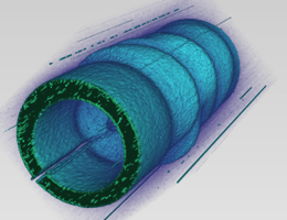
 A three-dimensional depiction of the spatial variation of the optical electromagnetic field around a microantenna following excitation with terahertz pulse. The optical field is mapped with the aid of electron pulses. (Illustration: Peter Baum)
The driving force behind all electronics is the temporally varying electromagnetic fields, whose polarities can change at extremely fast rates. Visualizing the action of these electromagnetic fields is a challenging task. The electron flow in electronic components like ‘field-effect’ transistors is controlled by these fields, making them responsible for data manipulation, flow, and storage in smartphones and computers.
However, gaining knowledge about the dynamics of field variation in such components is crucial for future developments in electronics. An important step towards this goal has been achieved by researchers at the Laboratory for Attosecond Physics, jointly operated by LMU and the Max Planck Institute for Quantum Optics (MPQ), who built an electron microscope capable of imaging high-frequency electromagnetic fields and tracing their ultrafast dynamics.
The new electron microscope exploits the ultrashort pulses of laser light. These pulses only last for a few femtoseconds. Bunches of electrons that consist of very few particles are generated using these laser pulses, and then temporally compressed by terahertz (1012 Hz) near-infrared radiation. The LMU and MPQ scientists, who are part of the research team in Ultrafast Electron Imaging, first demonstrated this method earlier this year in the Science journal, where they demonstrated the techniques capability to produce electron pulses, which are shorter than an optical half-cycle.
In the latest research, the physicists demonstrate the way of mapping high-frequency electromagnetic fields using these ultra-short electron pulses. The experiment involves directing the pulses onto a microantenna, which has just interacted with a precisely timed burst of terahertz radiation. The surface electrons in the microantenna are excited by the light pulse, generating an oscillating optical (electromagnetic) field in the near field of the target.
The electron pulses scatter when they are within the influence of the induced electromagnetic field around the microantenna. Their deflection pattern is then recorded. The spatial distribution, orientation, and temporal variation of the light released by the antenna can be reconstructed by the researchers based on the dispersion of the deflected electrons.
In order to visualize electromagnetic fields oscillating at optical frequencies, two important conditions must be met. The duration of each electron pulse, and the time it takes to pass through the region of interest, must both be less than a single oscillation period of the light field.
Dr. Peter Baum, LMU
The rate of propagation of the electron pulses used in this study is approximately equivalent to half the speed of light.
Using the principle of the electron microscope, the researchers have demonstrated the feasibility of precisely detecting and measuring even the smallest and most rapidly oscillating electromagnetic fields. This capability will enable the physicists to gain in-depth knowledge about the operation of transistors or optoelectronic switches at the microscopic level.
The sophisticated microscope can also be used for the advancement and examination of metamaterials, which are synthetic, patterned nanostructures with fundamentally different permeability and permittivity for electrical and magnetic fields, respectively, when compared to naturally occurring materials.
With these metamaterials, novel optical phenomena can be realized, which is otherwise not possible with traditional materials, paving the way for new opportunities in optics and optoelectronics. For example, it is possible to develop the fundamental building blocks to fabricate components for light-driven circuits and computers. This new method of characterizing electromagnetic waveforms using attosecond physics is a step towards future electronics.